Back Muscles Diagram Labeled - Back Muscles Diagram Labeled Vertebral | Wiring Diagram Database. It is also referred to as myopropulsive tissue. {label gallery} get some ideas to make labels for bottles, jars, packages, products, boxes or classroom activities for free. The deltoid, teres major, teres minor, infraspinatus, supraspinatus (not shown) and subscapularis muscles (not shown) all extend from the scapula to the humerus and act on the shoulder joint. Muscles of the back can be divided into superficial, intermediate, and deep group.since the all the back muscles originate in embryo (fetus) form by locations other than the back, muscles in the. These muscles are able to move the upper limb as they originate at the vertebral column and insert onto.
There are anterior muscles diagrams and posterior muscles diagrams. Human muscle system, the muscles of the human body that work the skeletal system, that are under voluntary control, and that are concerned with movement, posture, and balance. Human sacrum bone structure diagram, anatomical vector illustration labeled scheme with bone sections. Using the word bank, label the muscles shown in the front view on this free worksheet. {label gallery} get some ideas to make labels for bottles, jars, packages, products, boxes or classroom activities for free.
BACK MUSCLES ANATOMY - JACKED 4EVER from 2.bp.blogspot.com Studying these is an ideal first step before moving labeled diagram. Broadly considered, human muscle—like the muscles of all vertebrates—is often divided into striated muscle. Ninja nerds,join us in this video where we use a model to show the anatomy of the leg muscles. Illustration about bodybuilding and anatomy. Human muscle system, the muscles of the human body that work the skeletal system, that are under voluntary control, and that are concerned with movement, posture, and balance. Muscle anatomy quiz for anatomy and physiology! Learn vocabulary, terms and more with flashcards, games and other study tools. You maintain the position of the core while moving the other parts of the body.
It also covers some common conditions and injuries that can affect the.
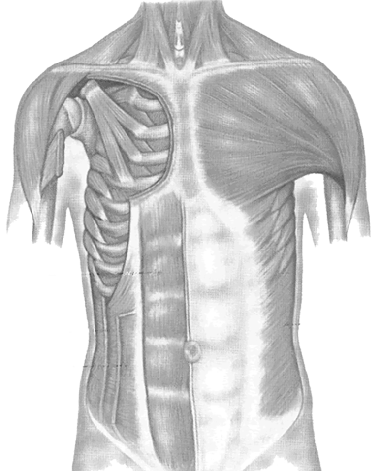
Download scientific diagram | anatomy of the intrinsic back muscles. Learn vocabulary, terms and more with flashcards, games and other study tools. Muscles also contribute to internal functions of the human body which include motion in the intestines and circulatory system. The human back extends from the buttocks to the posterior portion of the neck and shoulders. Related posts of muscles labeled front and back. Pancreas anatomical cross section model, vector illustration medical example. Tutorials and quizzes on the anatomy and actions of the back muscles (iliocostalis, longissimus, spinalis, multifidus, and quadratus lumborum), using interactive animations, diagrams, and illustrations. Back anatomy | all about the back muscles the back anatomy includes the latissimus dorsi, trapezius, erector spinae, rhomboid, and the teres major. Back to blank muscle diagram. Muscle tissue is a soft tissue that composes muscles in animal bodies, and gives rise to muscles' ability to contract. It is opposite from the chest, and the vertebral column runs the best way to strengthen back muscles is in a static position. The muscular system is responsible for movement in collaboration with the nervous system to form impulses for motion. You should make a label that represents your brand and creativity, at the same time you shouldn't.
Muscular system without labels muscular system diagram blank muscular system diagram with labels. {label gallery} get some ideas to make labels for bottles, jars, packages, products, boxes or classroom activities for free. It is also referred to as myopropulsive tissue. It is opposite from the chest, and the vertebral column runs the best way to strengthen back muscles is in a static position. These muscles are able to move the upper limb as they originate at the vertebral column and insert onto.
Muscles of the Back and Chest from www.biologycorner.com Related posts of back muscle diagrams labeled chest muscle diagram. Download scientific diagram | anatomy of the intrinsic back muscles. Studying these is an ideal first step before moving labeled diagram. Identify the muscle labeled as 1 in the diagram above There are anterior muscles diagrams and posterior muscles diagrams. Using the word bank, label the muscles shown in the front view on this free worksheet. Line diagram of axial section at the level of l3 (a); The deltoid, teres major, teres minor, infraspinatus, supraspinatus (not shown) and subscapularis muscles (not shown) all extend from the scapula to the humerus and act on the shoulder joint.
12 photos of the muscles labeled front and back.
It is opposite from the chest, and the vertebral column runs the best way to strengthen back muscles is in a static position. Tutorials and quizzes on the anatomy and actions of the back muscles (iliocostalis, longissimus, spinalis, multifidus, and quadratus lumborum), using interactive animations, diagrams, and illustrations. Muscle diagrams are a great way to get an overview of all of the muscles within a body region. Ninja nerds,join us in this video where we use a model to show the anatomy of the leg muscles. Muscles of the back can be divided into superficial, intermediate, and deep group.since the all the back muscles originate in embryo (fetus) form by locations other than the back, muscles in the. Use the location, shape and surrounding structures to. An easy and convenient way to make label is to generate some ideas first. Nerve root anatomical structure labeled cross section. Related posts of muscles labeled front and back. Related posts of back muscle diagrams labeled chest muscle diagram. Within this group of back muscles you will find the latissimus dorsi, the trapezius, levator scapulae and the rhomboids. Learn anatomy muscle labeling with free interactive flashcards. Muscle anatomy quiz for anatomy and physiology!
Nerve root anatomical structure labeled cross section. Torso diagram neck shoulder 3d illustration 3d rendering anatomical anatomy athlete back body bodybuilding bursa buttocks chart deltoid elbow fitness gluteus gluteus maximus gracilis health healthy human human anatomy 3d isolated on white joint label latissimus dorsi ligament lower back muscles. Back anatomy | all about the back muscles the back anatomy includes the latissimus dorsi, trapezius, erector spinae, rhomboid, and the teres major. Tutorials and quizzes on the anatomy and actions of the back muscles (iliocostalis, longissimus, spinalis, multifidus, and quadratus lumborum), using interactive animations, diagrams, and illustrations. Pancreas anatomical cross section model, vector illustration medical example.
Diagrams of Back Muscles | 101 Diagrams from www.101diagrams.com Many conditions and injuries can affect the back. Download scientific diagram | anatomy of the intrinsic back muscles. Muscle anatomy quiz for anatomy and physiology! This is opposed to other components or tissues in muscle such as tendons or perimysium. These muscles are able to move the upper limb as they originate at the vertebral column and insert onto. Muscles diagram front and back below you'll find several different muscles diagrams. Start studying back muscle labeling. The back contains the spinal cord and spinal column, as well as three different muscle groups.
Now label the diagram in your workbook!
The general action of the back muscles allows movement in the head, shoulders, arms, and the spine they are also involved in movement of the ribs which allows for respiratory function. There are anterior muscles diagrams and posterior muscles diagrams. Muscle anatomy quiz for anatomy and physiology! It is opposite from the chest, and the vertebral column runs the best way to strengthen back muscles is in a static position. Click on the labels below to find out more about your muscles. It also covers some common conditions and injuries that can affect the. Back to blank muscle diagram. Male muscular system, full anatomical body diagram with muscle scheme, vector illustration educational poster. The superficial back muscles are the muscles found just under the skin. Related posts of back muscle diagrams labeled chest muscle diagram. Many conditions and injuries can affect the back. 11.01.2020 · we are pleased to provide you with the picture named anatomy of back muscles diagram.we hope this picture anatomy of back muscles diagram can help you study and research. The deltoid, teres major, teres minor, infraspinatus, supraspinatus (not shown) and subscapularis muscles (not shown) all extend from the scapula to the humerus and act on the shoulder joint.
The superficial back muscles are the muscles found just under the skin back muscles diagram. Back to blank muscle diagram.
Share :
Post a Comment
for "Back Muscles Diagram Labeled - Back Muscles Diagram Labeled Vertebral | Wiring Diagram Database"


Post a Comment for "Back Muscles Diagram Labeled - Back Muscles Diagram Labeled Vertebral | Wiring Diagram Database"